PATIENT VIRTUALIZATION
The development of innovative technologies, together with their disruptive entry into daily clinical practice, has led to a profound reorganization and simplification of workflows.
Today, digital technology allows for complete patient virtualization in dentistry, as well as in general medicine.
Dentists are now able to gather all the patient's information at the appropriate times and in the appropriate manner, allowing the specialist to accurately approach the assessment and study of the clinical case. This is a true revolution, especially for complex cases and those requiring a multidisciplinary treatment plan.
Having a "virtual patient" means having a precise, accurate, and comprehensive overview of the initial clinical picture. This makes it possible to propose personalized and appropriate treatment options, which can be presented appropriately to the patient. This also allows for a two-way dialogue aimed at obtaining informed consent for treatment.
Finally, virtualization allows for improved diagnosis and treatment, as well as reestablishing and strengthening the doctor-patient relationship of trust. Technology thus lends itself to the service of security and transparency.
The case study – as it was called in a not-so-distant past – consisted of taking impressions and, in more complex cases, taking a facebow, together with the radiological and photographic examinations necessary for correct and in-depth planning.
A diagnostic wax-up proceeded with the simulation, on an articulator, of the prosthetic rehabilitation, while a photographic editing could be used for communication with the patient.
When rehabilitation simultaneously involves morphological, aesthetic and functional areas, the different planning elements certainly represent a less empirical starting point. Despite this, they are "disconnected from each other", not allowing for the full integration of information relating to the prosthetic project with the consequent evaluations of its impact on the dynamics or aesthetics of the work.[1]
Today, the term "patient virtualization" takes on a much broader and more comprehensive meaning. The ability to acquire static and dynamic three-dimensional files and the ability to "align" them with one another allows the creation of an avatar that features exactly the same physiognomic profiles and the same positioning of the dental arches as the aesthetic profile being examined.
This provides the specialist with an initial simulation of how the new prosthetic design will fit into the patient's aesthetics. With Itaka, this information can be integrated with an analysis of the patient's subjective chewing dynamics.
This is where IDI EVOLUTION's new digital platform, ALFRED, fits in. It standardizes the process of gathering the information needed for patient virtualization. The platform also assists and streamlines the workflow of the dentist-ASO-receptionist team: thanks to ALFRED, each team member can confidently carry out their work while also caring for the patient with the appropriate attention and empathy.
The philosophy that guides operational practice is divided into three elements: virtualize, design, and implement.
Using a technology like ALFRED—which can streamline and streamline the various operational phases—provides significant organizational advantages, as well as a significant reduction in waste in terms of materials, steps, and time. Furthermore, the platform allows for a more efficient and cost-effective approach. lean , more efficient “dental clinic system”.
ALFRED collects all patient information in a single silo: medical history, extra-oral examination, facial scan, clinical intra-oral examination, photographs, intra-oral scan of the dental arches, radiology (including CBCT), virtual implant-prosthetic design for guided surgery, information on the bone quality of the implant site, information on the integral-stability of the implant measured by the TMM3® surgical motor , registration of mandibular dynamics with ITAKA .
In recent times, there has been increasing discussion about digital technologies applied in dentistry and dental technology. However, only recently has it been possible to develop a prosthetic rehabilitation capable of including digital evaluation of chewing dynamics.
Gnathology, with all its aspects and schools of thought, still lives within the constraints created by the tools that are still used today to recreate, in all its complexity, a model of the stomatognathic system.
New technologies make it possible to record the dynamics of chewing, and these tracings are then aligned with patient models and inserted into a three-dimensional facial scan. This creates a precise anatomical model consisting of the dental arches, which contains information about the prosthetic rehabilitation to be performed, information about the soft tissues, and, above all, the precise dynamics of the models themselves. This allows for immediate identification of any interference or the congruence of the artifact and the recorded dynamic values during the digital modeling phase.
The following clinical case – created by Dr. Alessandro Galluzzi – is a clear example of the opportunities offered today by "patient virtualization" to respond to both aesthetic and functional needs, truly tailored to the patient.
Clinical case
The patient comes to the office asking for a reevaluation of the functional aspect of chewing and the aesthetics of the previously rehabilitated areas.
Clinical and gnathological examinations show a preservation of the VDO. Furthermore, masticatory dynamics are congruent, while the difficulties are attributable to the lack of elements in the distal regions. The fixed prosthesis applied is asymmetrical and has an inappropriate overjet.
The rehabilitation plan includes the insertion of osseointegrated implants to restore the dental abutments and the replacement of existing prostheses while respecting the patient's aesthetics and chewing function.
Patient virtualization begins with a TRIOS 3Shape intraoral scan, aligned with the CBCT by the IDI MAKERS digital dental technology department. Based on these models, the implant design is created, as well as the preparation of a support (fork) to be attached to the patient's lower arch for recording mandibular movements with ITAKA .
To complete the information acquisition, a scan of the patient's face is performed with different facial expressions ( RAYFACE ) and the mandibular movements of opening, closing and laterality in the rest position are detected, also including the chewing of a soft bolus in the measurements.
The three-dimensional model created allows for the creation of a temporary denture that falls within the aesthetic parameters defined and controlled through the superposition of scans of the patient's face and the functional parameters identified and recorded with ITAKA .
Already from the application of the temporary prosthesis it is possible to appreciate how the collection of such a precise and detailed set of information allows for a reduction in both aesthetic and occlusal corrective interventions.
The final impression is always taken using a digital technique and, subsequently, a new recording of the masticatory dynamics is carried out after a conditioning period with the temporary prosthesis.
The new findings take on a broader significance: in addition to providing a new dynamic model on which to design the definitive prosthesis, they allow for a reassessment of the suitability of the applied temporary prosthesis.
Upon delivery of the definitive prosthesis, always made by the IDI MAKERS department in 1200 mpa zirconia, no aesthetic or functional adjustments were necessary.
While chewing, the patient found the solution comfortable, free of interference or pre-contact, confirming that innovation and technology are creating a new safety and quality standard, even in cases of extensive and complex rehabilitation.

Fig. 1 Initial clinical situation of the patient

Fig. 2 Facial scan

Fig. 3 4D prosthetic design in mandibular dynamics with ITAKA movement paths

Fig. 4 The virtual implant project for guided surgery

Fig. 5 Final prosthetic rehabilitation in zirconia: upper arch

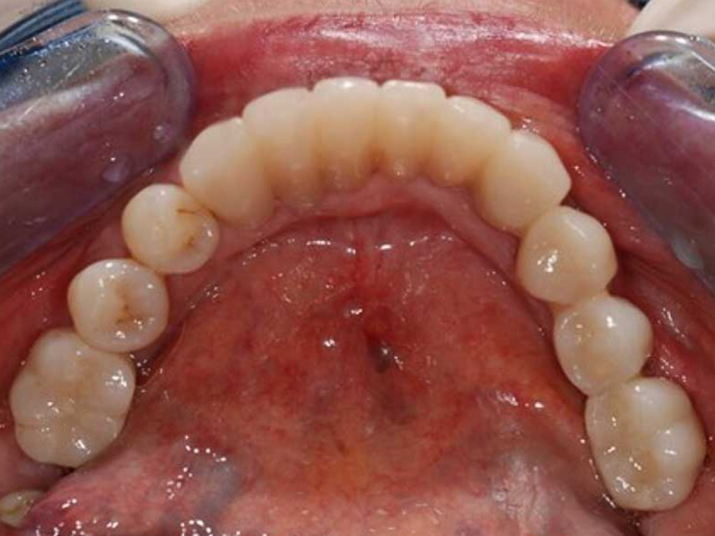
Fig. 6 Final prosthetic rehabilitation in zirconia: lower arch

Fig. 7 Final aesthetics of prosthetic rehabilitation
Read the article on Odontoiatria33.

Share: